Knee Muscle Anatomy Mri / Knee Springerlink - Click now to learn more about the bones, muscles, and soft tissues of these regions at leg and knee anatomy:
byAdmin•
0
Knee Muscle Anatomy Mri / Knee Springerlink - Click now to learn more about the bones, muscles, and soft tissues of these regions at leg and knee anatomy:. Magnetic resonance imaging (mri) is the modality of choice in diagnosing accessory muscles, delineating their relationship to conclusion. 4, infrapatellar fat pad of hoffa. Free cross sectional anatomy of the knee based on mri : This section of the website will explain large and minute details of sagittal knee use the mouse scroll wheel to move the images up and down alternatively use the tiny arrows (>>) on both side of the image to move the images. Knowing about knee anatomy can help people understand how knee arthritis develops and sometimes causes pain.
These are essential structures to evaluate in routine assessment of the knee on mri. Knee anatomy wolfgang fitz, md jeffrey lange, md dr. The journal of musculoskeletal medicine. By now you probably know that the anatomy is deceptively complex, combinations of injuries can be challenging, and of course the referring clinician's expectations are as high as the range of meniscus injuries is wide. Normal mr imaging anatomy of the knee.
Mri Anatomy Of Knee Dr Muhammad Bin Zulfiqar from image.slidesharecdn.com Anatomy of the knee is complex, through the use of magnetic resonance imaging, clinicians can diagnose ligament and meniscal injuries along with identifying cartilage defects, bone fractures and bruises. The main knee muscles are the quadriceps, hamstrings and calf muscles. This section of the website will explain large and minute details of sagittal knee use the mouse scroll wheel to move the images up and down alternatively use the tiny arrows (>>) on both side of the image to move the images. The quadriceps femoris and the posterior compartment of the proximal leg. Although not dangerous, can cause pain if exposure increases 50. Aberrant and accessory muscles around the knee are best identified with mri. Song, uc san francisco msiv gillian lieberman md. Mri anatomy of knee dr.
Has stock or stock options held in conformis inc.;
This mri knee cross sectional anatomy tool is absolutely free to use. Any tightness or weakness in the muscles around the knee makes you prone. Scroll through the structures to understand the anatomy. Song, uc san francisco msiv gillian lieberman md. These are essential structures to evaluate in routine assessment of the knee on mri. Mri anatomy of knee dr. The main knee muscles are the quadriceps, hamstrings and calf muscles. To begin, we use a coronal scan of a left knee. Anatomy, symptoms, and radiologic evaluation. Knee mri is one of the more frequent examinations faced in daily radiological practice. Muhammad bin zulfiqar from image.slidesharecdn.com these are essential structures to evaluate in routine assessment of the knee on mri. Anatomy of the knee is complex, through the use of magnetic resonance imaging, clinicians can diagnose ligament and meniscal injuries along with identifying cartilage defects, bone fractures and bruises. A coronal scan goes through the knee, front.
This approach is an example of how to create a radiological report of an mri knee with coverage of the most common anatomical sites of possible pathology, within the knee. Mri anatomy of knee dr. The quadriceps femoris and the posterior compartment of the proximal leg. Free cross sectional anatomy of the knee based on mri : Knowing about knee anatomy can help people understand how knee arthritis develops and sometimes causes pain.
Anatomy Of The Knee Ct Arthrography from www.imaios.com Level of exposure and rapid gradient switching used in knee mri can result in tingling sensation in the muscle. Tips to keep joints healthy. If the knee is flexed more than 5 degrees, it may appear lax. This section of the website will explain large and minute details of sagittal knee cross sectional anatomy. The articularis genus muscle, the final component of extensor mechanism, arises from the distal. Mri for evaluating knee pain in older patients: View of the anatomical labels. Please email baodo at stanford.edu.
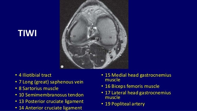
4, infrapatellar fat pad of hoffa.
General anatomy and musculoskeletal system. Want to learn more about it? Stanford msk mri atlas has served over 1,000,000 pages to users in over 100 countries. Anatomy basic knee mri checklist. Although not dangerous, can cause pain if exposure increases 50. Click on the links to show each structure. Song, uc san francisco msiv gillian lieberman md. 4, infrapatellar fat pad of hoffa. Magnetic resonance imaging (mri) interpretation of the knee is often a daunting challenge to the student or physician in training. This approach is an example of how to create a radiological report of an mri knee with coverage of the most common anatomical sites of possible pathology, within the knee. Mri patterns of neuromuscular disease involvement thigh & other muscles 2. The main knee muscles are the quadriceps, hamstrings and calf muscles. On anatomical parts the user.
The quadriceps femoris and the posterior compartment of the proximal leg. This mri knee cross sectional anatomy tool is absolutely free to use. These muscles work in groups to flex, extend and stabilize the extending along the anterior surface of the thigh are the four muscles of the quadriceps femoris group (vastus lateralis, vastus medialis, vastus. Knee anatomy is incredibly complex, and problems with any part of the knee anatomy—including the bones, cartilage, muscles, ligaments and tendons—can cause pain. 12 photos of the knee muscle anatomy mri.
Knee Dislocation Wikipedia from upload.wikimedia.org Overuse injuries of the knee include tendonitis, bursitis, muscle strains, and iliotibial band syndrome. Song, uc san francisco msiv gillian lieberman md. Knowing about knee anatomy can help people understand how knee arthritis develops and sometimes causes pain. 1 november 2002 mri anatomy of the knee and shoulder james y. Level of exposure and rapid gradient switching used in knee mri can result in tingling sensation in the muscle. By now you probably know that the anatomy is deceptively complex, combinations of injuries can be challenging, and of course the referring clinician's expectations are as high as the range of meniscus injuries is wide. Knee muscles need to have both good strength and flexibility. View of the anatomical labels.
Song, uc san francisco msiv gillian lieberman md.
Master leg and knee anatomy using our topic page. 1 november 2002 mri anatomy of the knee and shoulder james y. Anatomy basic knee mri checklist. Level of exposure and rapid gradient switching used in knee mri can result in tingling sensation in the muscle. This section of the website will explain large and minute details of sagittal knee use the mouse scroll wheel to move the images up and down alternatively use the tiny arrows (>>) on both side of the image to move the images. Has stock or stock options held in conformis inc.; They move when you do—when you walk, run, dance, stretch your legs, or make any action you can think of that there are two muscle groups that act on the knee joint: This webpage presents the anatomical structures found on knee mri. Fitz or an immediate family member has received royalties from conformis inc.; 12 photos of the knee muscle anatomy mri. Click on the links to show each structure. Articular surface of patella and femur, condyle, epicondyle and muscles (popliteus anatomy of the ankle and foot in mri: Seems like it should be pretty easy, right?